Your data on MRCVSonline
The nature of the services provided by Vision Media means that we might obtain certain information about you.
Please read our Data Protection and Privacy Policy for details.

In addition, (with your consent) some parts of our website may store a 'cookie' in your browser for the purposes of
functionality or performance monitoring.
Click here to manage your settings.
If you would like to forward this story on to a friend, simply fill in the form below and click send.

Your friend's email:
Your email:
Your name:
 
 
Send Cancel

Dick Vet study to compare equine MRI and CT scans
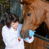
Vets Sarah Taylor and Padraig Kelly of the Dick Vet Equine Hospital.
Research will make use of newly installed CT scanner.

A new three-year study at the University of Edinburgh’s Dick Vet Equine Hospital will compare the use of MRI and CT scans for horses with lameness and foot problems.

The study has been made possible following the delivery
earlier this month of a new CT scanner for scanning the distal limbs of sedated standing horses. The equipment has been housed in a purpose-built room at the hospital and will help the veterinary team in their diagnostic work.

During the next three years, horses referred to the hospital for an MRI scan will undergo a CT scan beforehand, at no extra cost to the client. The CT scan will allow the veterinary team to check for metal clenches in the hoof wall before the MRI to avoid the metal migrating in the MRI and causing injuries, replacing the use of X-rays.

Researchers will also look at whether using an MRI or CT scan, or both, is necessary for diagnosing different equine foot conditions. They will use anonymised images to compare the sensitivity and specificity of the two scanning methods for navicular syndrome and coffin joint osteoarthritis.

The findings will help the veterinary team decide which scanner to use when diagnosing patients with equine distal limb problems to minimise over-imaging.

Padraig Kelly, head of the Dick Vet Equine Hospital, said: “We are excited to offer both standing MRI and CT scans at no extra cost to our clients. This will significantly aid the diagnosis of lameness of our patients.

“Having both imaging modalities will also provide an excellent opportunity to do some sensitivity and specificity studies to determine whether CT or MRI is better for detecting different injuries in a horse's foot.”

Image © Shutterstock

Become a member or log in to add this story to your CPD history

Prof Joanne Webster elected as Fellow of the Royal Society

News Story 1
 Joanne Webster, a professor of parasitic diseases at the RVC, has been elected as a Fellow of the Royal Society (FRS).

An infectious disease expert, Prof Webster is known for promoting a One Health approach to disease control.

She completed her doctoral research in zoonotic disease and parasite-host interactions, and has since earned widespread recognition for contributions to parasitology and global health.

Prof Webster said: "I am truly honoured, and somewhat stunned, to be recognised alongside such an exceptional group of scientists." 

Click here for more...
News Shorts
Germany FMD import restrictions eased

The UK government has lifted the import restrictions placed on FMD-susceptible commodities from Germany.

The decision comes after the country was recognised as foot-and-mouth disease free without vaccination on 14 May.

Imports of FMD-susceptible animals and their by-products from Germany were originally banned, after the country reported a case of FMD near Brandenburg in January. In March, the UK government permitted imports from outside of the outbreak zone.

Germany will now be able to import FMD-susceptible animals and their by-products into the UK, providing they meet other import conditions.

The decision follows rigorous technical assessment of measures in Germany. Defra says it will not hesitate respond to FMD outbreaks.