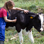
Prion research used imaging process called electron cryomicroscopy
The structure of the infectious agent that causes bovine spongiform encephalopathy (BSE) has been identified by researchers at the University of Alberta.
BSE, commonly known as "mad cow disease", is caused by a misfolded protein called a prion. Until now, all attempts to shed light on the structure of the protein have failed due to its tendency to clump together.
Writing in the journal PLOS Pathogens, the team describe how they obtained a very simple, preliminary idea of the structure using an imaging process called electron cryomicroscopy. The researchers say the structure argues against existing theories of prion conversion and suggests how the process might actually work.
"The recent advances in electron cryomicroscopy technology are certainly a breakthrough," explains co-principal investigator Holger Wille. "We know the structure of the normal cellular form of the protein, but we know very little about the infectious prion protein and how it propagates. The use of these high-powered microscopes has finally given us some clarity."
In the study, the team used electron cryomicroscopy to collect thousands of high-resolution micrographs. From these, the team extracted the best images to build a three-dimensional model for the structure of the infectious prion protein. The study suggests how infectious prions replicate by converting non-infectious, cellular versions into copies of themselves.
"It is not an atomistic model, so we cannot say which position the atoms are in," says Wille. "But this is something we hope to do in the future."
Looking ahead, the researchers wish to study the structure in more depth. The study used model system prions, but they are now using prions that infect cows (BSE), wild animals (chronic wasting disease) and humans (Crreutzfeldt-Jakob disease).
"Ultimately, if we know how the prion propagates, we could come up with clinical interventions to treat or prevent disease," adds Wille.


 HMRC has invited feedback to its communications regarding the employment status of locum vets and vet nurses.