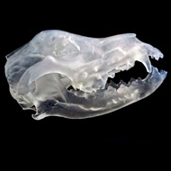
Design project enhances treatment options for animals
US vets are using 3D printed models of fractured and deformed animal bones for teaching and planning surgeries.
The 3D prints have been developed as part of a collaborative project between product design student Kelsey Catinado, professor Dustin Headley, and Kansas State University's College of Veterinary Medicine.
The printing process retains and enhances the important information found on a scan that a doctor or vet needs in order to make diagnosis.
Walter Renberg, orthopaedic surgeon and head of small animal surgery at the college's Veterinary Health Center, said the 3D models are proving beneficial in a variety of ways:
"It helps us with a couple of things clinically, particularly with bone deformities, which can be difficult to reconstruct with a CT scan. For example, when planning a surgery to correct a deformity or even determining whether such a surgery is necessary, the model can help us determine the right surgical approach or come up with less expensive alternatives to certain procedures."
Earlier this summer, a 3D print made of a dog's malformed tibia did just that.
Renberg added: "I thought we would have to do an expensive reconstruction that the client probably couldn't afford, but the 3-D modelling gave us a better understanding of the problem and we came up with a less invasive and less expensive route."
For the project, Castinado used digital files of CT scans provided by the Veterinary Health Centre. As each file contains small, chopped-up fragments of bone, Castinado used 3D modelling software to bring all the pieces together. She then removed all the extra fragments that are attached, so that when it is printed in 3D, it looks like a bone.
As well as helping to plan surgeries and find more cost-effective treatment options, the 3D printed models are also being used by vets as teaching aids.
"From a clinical standpoint, we can use the 3D models with clients to explain procedures," Renberg said. "It can be easier to show them a model than a CT scan."
Work is ongoing to to see if 3D printing could be used in other ways, such as exploring soft tissues in 3D at scale.
Image (C) Kansas State University.


 The Veterinary Medicines Directorate (VMD) is inviting applications from veterinary students to attend a one-week extramural studies (EMS) placement in July 2026.