
The new technology could support tailored cancer care for cats and dogs.
Researchers at the Royal (Dick) School of Veterinary Studies are utilising advanced fluorescent imaging to detect potential sites of tumour spread in dogs and cats.
The camera-based technique, known as Near-Infrared Fluorescence (NIRF) imaging, enables soft tissue surgeons at the Hospital for Small Animals to locate and remove tiny lymph nodes that may affect how cancer spreads.
It uses a harmless green dye, which is injected into the tumour while the animal remains under general anaesthetic. The dye appears bright green under a specialised near-infrared camera and is easily visible as it flows along the lymphatic pathways and into the lymph nodes.
The skin and tissues appear normal to the naked eye, so the green glow can help the surgeon detect lymph nodes that must be removed with accuracy and confidence. It can also help the surgeon confirm how many lymph nodes to remove.
Researchers note the method is safer for patients and enables vets to determine the stage of the cancer more accurately. This can also help vets support tailored cancer care for pets.
“We are delighted to have this technology available that allows us to work with our colleagues in Oncology to strengthen the accuracy and completeness of staging, and support tailored oncology decision-making,” says Dr Kathryn Pratschke, a specialist in soft tissue surgery at the Hospital for Small Animals. “This will help improve outcomes for patients referred to us for treatment of cancer.”
Professor Laura Blackwood, head of oncology service, Hospital for Small Animals, added: “Having NIRF available significantly improves the management of patients with tumours with high risk of lymph node spread. It's great to have it available for our patients.”
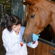
Image (C) Casey Kidd/Shutterstock.com

 The latest